Mobile Lab in Stanford Operating Rooms is key to Precision Health
Mar 14 2019
Posted In:
20/20 Blog
Palo Alto, CA — Vinit Mahajan, M.D., Ph.D., Associate Professor of Ophthalmology in Stanford University School of Medicine’s Department of Ophthalmology, believes that eye tissue discarded during surgery holds the clues researchers need to prevent and cure both common and rare eye diseases. These tissues are extremely valuable for molecular experiments aimed at finding new diagnostic tests and novel treatments.
One of the obstacles to preserving tissues from the operating room is the physical distance between medical facilities and research buildings. With this in mind, Dr. Mahajan established collaborations between surgeons and research scientists by creating a Mobile Operating Room Laboratory Interface (MORLI) that links the operating room with the research laboratory. The MORLI is the cornerstone of the Stanford Biorepository of Eye & Surgical Tissue (BEST) in the Department of Ophthalmology, an initiative that was spearheaded by Dr. Mahajan and Prithvi Mruthyunjaya M.D., Associate Professor of Ophthalmology. Since 2018, this departmental biorepository has been built and managed by clinical research coordinator Teja Chemudupati with the cooperation of the operating room team at Byers Eye Institute.
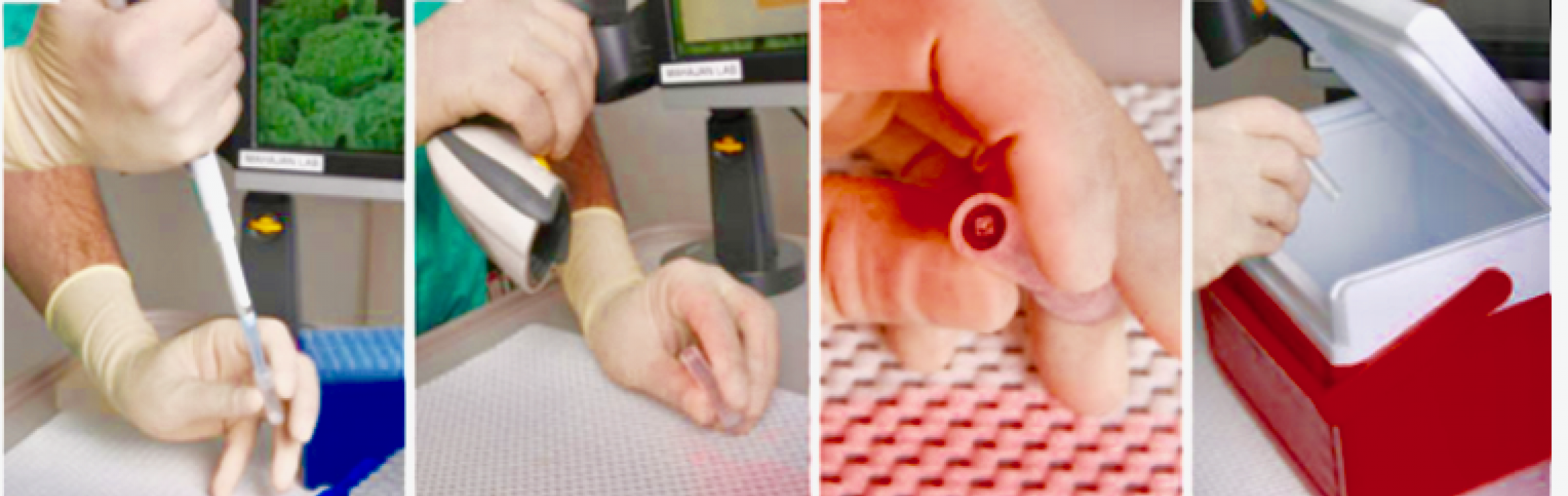
The MORLI is a laboratory on wheels designed to simulate a laboratory bench with all the required instruments and devices. This makes it possible for the surgical team to immediately process specimens in the operating room. The MORLI consists of a computer and bar code scanner for logging patient and sample information as well as lab supplies necessary for processing and preserving tissue samples. The unit holds pipettors, disposable pipette tips, centrifuge tubes, a centrifuge, a small liquid nitrogen dewer or other cryofreezer, and a binocular dissecting microscope.
Dr. Mahajan and his team found that on-site processing helped to maintain specimen integrity, since it was not possible to immediately or easily transfer specimens to the lab during or between surgery cases. The specimens are flash frozen and placed into a cooler which is then transported to a -80 laboratory freezer biorepository for long term storage.
Chemudupati explained, “Flash freezing tissue allows us to preserve molecules that would otherwise degenerate in minutes. The common practice of fixing tissue in wax is no longer the optimum method of preservation because it prevents researchers from using modern tools like sequencing the genome, proteomics, and metabolomics that analyze molecules."
Surgical staff and researchers use the MORLI’s computer to access a database with a user interface designed to accommodate sample collection and archival data. The user interface is designed for efficient entry of patient demographics, phenotype data, and sample features in a format familiar to surgeons and clinical personnel alike.
The MORLI system allows surgeons and a team of scrub techs, nurses, and clinical research coordinators to catalog, process, and store tissues using standardized procedures that maintain the integrity of tissue samples. Without standardized procedures, the composition of collected tissue can change leading to experimental artifacts rather than the disease mechanism being studied.
At the time of surgery, Stanford glaucoma, oculoplastic, retina, and cataract surgeons ask patients if they would like to donate the tissue that is normally excised and discarded during their procedure to an ophthalmology biorepository. Patient consents and deidentification protection measures allow researchers to collect and maintain tissue samples linked to important clinical disease information.
To date, thousands of bar-coded samples have been organized in a biorepository where they can be easily retrieved for current and future investigations of disease mechanisms, therapeutics, and diagnostics. A custom database streamlines tissue sample logging and retrieval, and it is linked to retrospective and prospective clinical data (all IRB approved). The database system has made multiple or rare specimens easy to locate.
Collected samples can be shared by researchers at Stanford and potentially with scientific collaborators across the country and around the world. Sample data shared with researchers outside the university are deidentified and require collaborators to work with Dr. Mahajan’s research team in order to access clinical data relevant to the study. Key collaborators in the BEST initiative include Rohit Gupta, the Executive Director of Clinical Research Services (CTRU) & Biobank, and his team who work to process and store blood draw samples collected from patients.
Important discoveries in eye cancers, rare diseases, and autoimmune diseases have already been made using tissues from the ophthalmology biorepository. The MORLI ensures that large collaborative projects in the future will have access to samples that are collected, processed, and stored in a consistent manner.
Mruthyunjaya said, “By replicating aspects of the lab in the operating room with the MORLI, we have provided a system for creating an efficient biorepository of human eye tissue samples, which can be applied to any type of biological sample collection.”