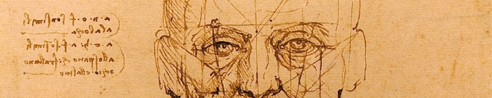
In his notebooks Leonardo da Vinci used his artistic skill not only for representation, but also as a tool for understanding and learning. His scientific drawings reveal incredible insight and an unquenchable curiosity for anatomy, civil engineering, optics and hydrodynamics. During his life, the sciences and the arts were not exclusive, and Da Vinci blended the two beautifully as he produced and maintained his notebooks on a daily basis.
The study of vitreoretinal surgical disease lends itself to Da Vinci’s visual style of exploration and learning. Despite advances in wide field digital imaging, it is still not possible to take complete fundus images in the preoperative, intraoperative, and postoperative stages. The events during and surrounding surgery can still be best seen, understood, and emphasized through hand drawings. Vitreoretinal fellows at the Univeristy of Iowa record surgical cases in a notebook that would have received da Vinci’s approval.
Surgeons enter a colored fundus drawing showing the pathology at the beginning of the case. At the case conclusion, the procedures that were preformed are overlaid on the original drawing. Annotations and labels are made around the drawing. An example is shown below.
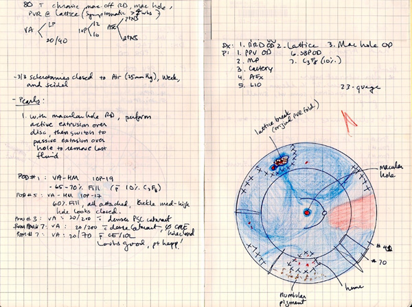
On the facing page, a brief historical note and preoperative exam findings are recorded. Surgical pearls, complications, and intraoperative events are described. Equally important, the postoperative examinations, events, and an occasional drawing are listed.
After recording a few cases like this, several benefits become apparent. First, it is much faster and more informative to review the case through a color drawing than reading highly standard operative notes. Second, keeping track of the postoperative outcomes is incredibly informative. Did the retina stay attached? How long did it take for the vision to stabilize? How long did it take for the pressure to return to normal? Where did tractional membranes develop? Finally, the fundus drawings and notes make it simple to share and discuss cases with colleagues.
Over time, the cases are indexed. That way, similar cases can be reviewed together. What technique worked best for giant retinal tears? How long does a combined case take? What vitrectomy settings were most efficient? Which suture technique worked best?
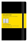
The Moleskine Notebook, with its simplicity, durability and portable size, is the perfect surgeon’s companion. It has been in use since the 1800s by famous artists such as Matisse, Van Gogh, Hemingway, and Picasso. The grid paper found in the soft covered squared Moleskine makes it easy to draw accurate images. Drafting templates with circles are used to draw the fundus outline and buckle elements. Colored pencils detail findings using the following color code:
| Element | Color |
|---|---|
| Retina, attached | Red (light) or white |
| Subretinal fluid | Blue, (hatched blue for retinoschesis) |
| Retinal Break | Red surrounded by blue outline |
| Retinal Hemorrhage | Red (dark) |
| Vitreous Hemorrhage, media opacities | Green |
| Choroidal and RPE hyper-pigmentation | Brown |
| Neovascularization | Red or purple (flat), orange (elevated) |
| Retinal Edema, Exudates, Drusen, Chorioretinitis | Yellow |
| Laser, cryotherapy | Black "X", back "X" inside a circle |
| Lattice | Black cross-hatch (blue cross hatch if detached retina) |