
Palo Alto, CA — The eyes may be a window into the soul, but Stanford research scientists are now showing the eyes may also be a window into overall human health.
In a few drops of eye fluid, the Mahajan Lab can reveal the same molecular data as tissue biopsies, opening a pathway to study fluids from the brain, lungs, kidney, and other organs that cannot be biopsied without causing irrevocable damage. Up until now, molecular details of these organs, including the eye, were best studied postmortem.
The team integrated microvolume liquid biopsy proteomics, single-cell transcriptomics and artificial intelligence to create a powerful tool to examine disease mechanisms at the cell level in living humans. They refer to this approach as TEMPO (Tracing Expression of Multiple Protein Origins).
Their manuscript, "Liquid biopsy proteomics combined with AI identifies cellular drivers of eye aging and disease in vivo" was recently published in Cell.
Vinit Mahajan M.D., Ph.D., Stanford professor and vice chair of ophthalmology research and director of the Molecular Surgery Program said, “Eye fluid protein concentration is about 400 times lower than in plasma, but an enhanced proteomics approach was a game changer. We detected almost 6000 proteins, ten times higher than had been measured before. And combining that with single cell transcriptomics gave us an overview of cell-specific gene expression in the eye.”
TEMPO allowed the Mahajan team to trace almost 6,000 proteins to specific normal and diseased cells in the eye. It revealed that the cellular drivers of diabetic retinopathy switch with disease stage. And surprisingly, it picked up proteins associated with Parkinson's Disease, showing that brain disease proteins are also expressed in the eye.
This information will be crucial for monitoring the progression of disease and testing novel therapies. It may also explain why current treatments are failing; they are not targeting the correct proteins.
Mahajan said, “Traditionally eye fluid obtained during cataract and retina surgeries is thrown away, but I had a hunch that eye fluid could hold molecular information that researchers could use in tracing eye disease and developing new therapies.”
Mahajan’s hunch was correct. And with four million cataracts performed every year in the US alone, researchers have ample access to this fluid.
Using TEMPO researchers found that approximately ninety percent of the proteins found in the vitreous, the gel like liquid attached to the retina, can be found in the aqueous humor, a water-like fluid in the front chamber of the eye.
Mahajan said, “This finding is especially important because it is safer and easier to collect aqueous humor fluid during cataract surgery or in the clinic compared to collecting vitreous fluid.”
Lab member Julian Wolf, M.D., an expert in big data analytics, took the TEMPO findings one step further by creating an AI platform that predicts the global and cellular molecular age of diseased eyes. AI modeling showed that the age of diseased eyes ranged from 12 to 28 years older than chronological age depending on the type and stage of the eye disease.
Dr. Wolf said, “These findings demonstrate that our organs are aging at different rates. The use of targeted anti-aging drugs could be the next step in preventative, precision medicine. For example, we found that the eyes of diabetic patients show advanced aging even when symptoms were absent. It is possible that we could prevent or delay symptoms with anti-aging drugs.”
TEMPO could usher in a whole new way for researchers to uncover the molecular mysteries of disease and aging. And it could provide specific information necessary to develop treatment and prevention plans based on an individual’s molecular health.
Mahajan lab research findings using eye fluid is so important to uncovering the molecular basis for eye disease that Stanford Ophthalmology invested in creating a biobank system to collect eye fluids that seamlessly connects the operating room with the laboratory. This resource allows surgeons and scientists to explore the next generation of diagnostics and therapeutics.
The team explains their study in a video abstract.
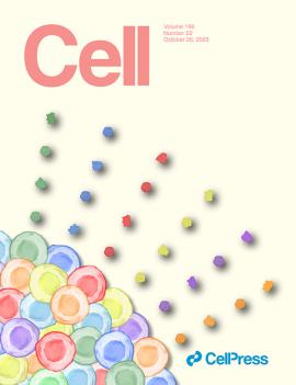
Artwork by Julian Wolf, Vinit Mahajan, and Antoine Dufour won the Cell cover contest. Inspired by the pointillism paintings of Georges Seurat, who deconstructed light into colored dots that our minds reconstruct into images, this cover depicts colored protein dots emerging from their like-colored cells. The TEMPO tool deconstructs human eye fluids into a proteome and reconstructs that data with single-cell gene expression.
Cell editors also selected the paper as a “featured article” for the October 26 issue.