ILM Technique Closes Macular Holes
Aug 9 2019
Posted In:
20/20 Blog
Palo Alto, CA – As the eye ages, vision problems increase, making it harder to do everyday activities. When an older patient comes in with blurred or distorted central vision, one of the causes an ophthalmologist will check for is a macular hole. Macular holes can develop as the aging vitreous, the clear gel that supports the eye, shrinks and pulls away from the retina. A macular hole can cause vision problems that affect reading, driving, and seeing fine detail.
Stanford University ophthalmologist Vinit Mahajan M.D., Ph.D., vitreoretinal surgeon and Vice Chair of Ophthalmology, said, “Macular holes are fairly common, and they can be associated with a number of eye conditions such as vitreous degeneration, severe nearsightedness, macular pucker, diabetic eye disease, detached retina, and eye injury.”
The most common treatment is a vitrectomy surgery where the internal limiting membrane (ILM) is peeled away to take the tension off of the retina and then the vitreous gel is replaced with a temporary gas or oil bubble. In some cases, this surgical technique may have some drawbacks. Mahajan has addressed these drawbacks with an alternative surgical method where an ILM peel is replaced by an abrasion of the ILM with a special surgical instrument, a diamond-dusted membrane scraper, at the time of vitrectomy.
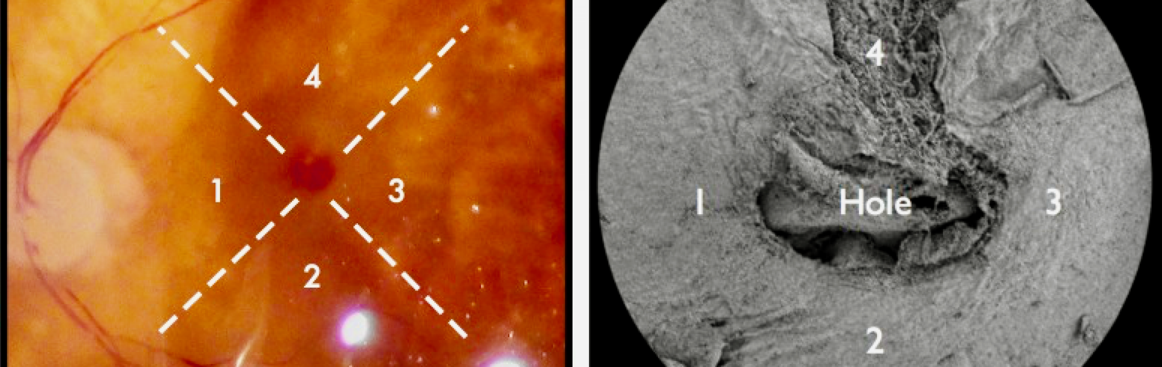
In laboratory studies, Mahajan’s research team studied the effect of the membrane scraper on human donor eyes. They found that the abrasion removed the fibrotic cells and “scar” tissue from the surface of the retina. At the same time, this method avoided damaging the underling axons of the retinal ganglion cells.
To determine the effectiveness of this technique in closing macular holes in patients, the surgical team reviewed 100 consecutive cases of macular hole surgery with ILM abrasion. This ILM abrasion technique achieved high rates of macular hole closure while preserving intrinsic foveal tissues and eliminating the need to use potentially toxic dyes to peel the thin membrane during vitrectomy surgery.
A few studies have suggested that indocyanine green, the chemical used in the dye most commonly used during vitrectomy, may cause toxicity to the retinal pigment epithelium (RPE) and neurosensory retina, as well as optic nerve atrophy in some cases. Different dyes are being explored for use. Although retina surgeons use dyes to help close macular holes, the research supports the use of an alternative method that may benefit patients.
To read more, click on the PDFs below:
Effect of Internal Limiting Membrane Abrasion on Retinal Tissues in Macular Holes
Macular Hole Closure With Internal Limiting Membrane Abrasion Technique